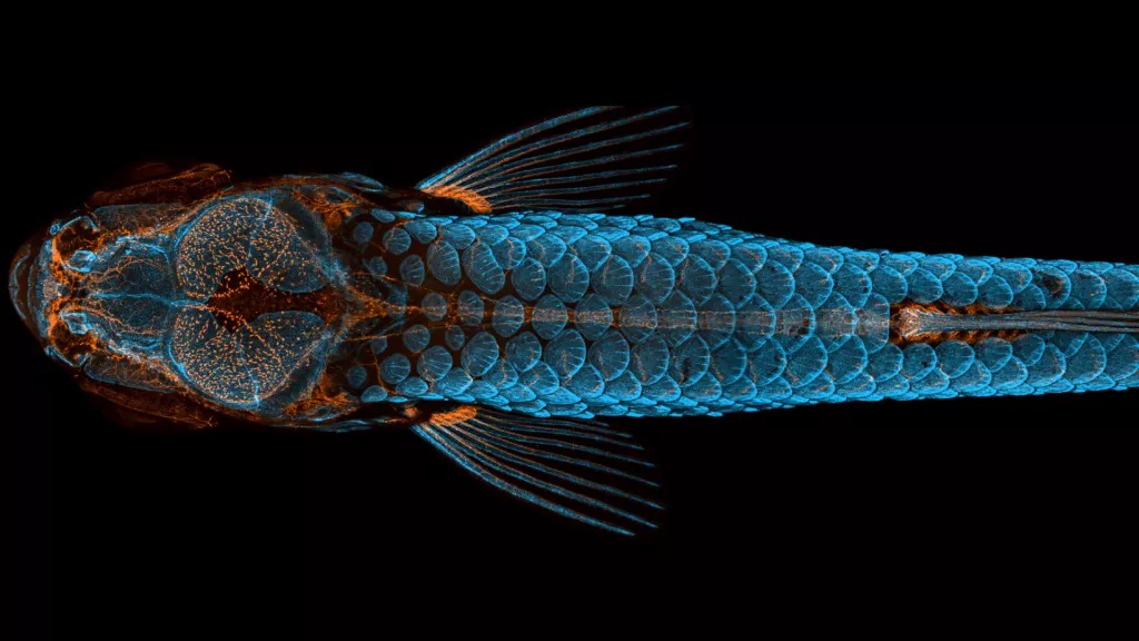
A vibrant, blue-and-orange hued photo highlighting the tiny vessels in and around the brain of a young zebrafish won first place in the Nikon Small World photography contest, an annual competition showcasing photos of our world at the microscopic level, Nikon representatives announced today (Oct. 13).
In this jaw-dropping image, lymphatic vessels in the fish’s brain glow orange, as do branching tendrils extending throughout its body in the complex network of the lymphatic system, which rids the body of waste. Offsetting the delicate orange threads are luminous blue patterns — the fish’s scales and bones.
Daniel Castranova, Brant Weinstein and Bakary Samasa, researchers at the National Institutes of Health (NIH) captured the prizewinning photo using confocal microscopy, an optical imaging technique. Castranova stitched together more than 350 images to achieve the final result, which contest representatives labeled “stunning” in a statement.
In zebrafish, the lymphatic vessels and bones express fluorescent proteins in different parts of the spectrum. Researchers then colorize illuminated regions in the images to differentiate between the different proteins, Castranova told Live Science.
To image the living zebrafish, Castranova and his colleagues anesthetized the animal and placed it in a pliable gel called agarose to hold the fish still for the photos, covering the zebrafish with water so that it could breathe.
“We can image them, and then peel them out of the agarose and revive them,” Castranova said. The researchers photographed this particular zebrafish during several stages of its growth, creating a time series to show where the lymphatic vessels in the brain come from and how they develop.
Lymphatic vessels were first discovered in mammal brains in 2015, and they may help the brain flush out toxins, according to the NIH. This zebrafish image is part of an investigation demonstrating for the first time that fish also have these vessels in their brains, he explained.
Second place went to a composite image showing embryo development in a clownfish (Amphiprion percula), in a sequence taken over four days by nature photographer Daniel Knop. The first egg on the far left was freshly fertilized, while the egg at the end of the line on the right was just hours away from hatching, according to the contest statement. The third-place winner, a highly colorful close-up of a freshwater snail’s tongue, was captured by Igor Siwanowicz, a biochemist and neurobiologist at the Max Planck Institute in Munich, Germany.
Minuscule marvels
For 46 years, Nikon’s contest has celebrated photographers and scientists who use microscopes and camera lenses to capture minuscule marvels. Standout images in previous years included a rainbow-colored turtle embryo; clusters of shimmering, sequin-like scales around a beetle’s eye; and a glowering embryonic fish face, to name just a few.
This year, winners were selected from over 2,000 entries, representing researchers and microscopy artists from 90 countries around the world. Judges evaluated submissions based on their artistic vision, originality, technical expertise and scientific context, according to the statement.
“First of all, the image has to be striking — it has to be aesthetically interesting or pleasing,” said contest judge Dylan Burnette, an assistant professor in the Department of Cell and Developmental Biology at the Vanderbilt University School of Medicine in Nashville, Tennessee.
“An interesting scientific angle is also required to push an image up to the top 20,” Burnette told Live Science. The clarity and beauty of Castranova’s image — as well as its scientific significance — instantly caught the judges’ attention, Burnette said.
“It was one of the only images that every one of the judges agreed on,” he said. “Its first-place position was pretty clear almost immediately.”


